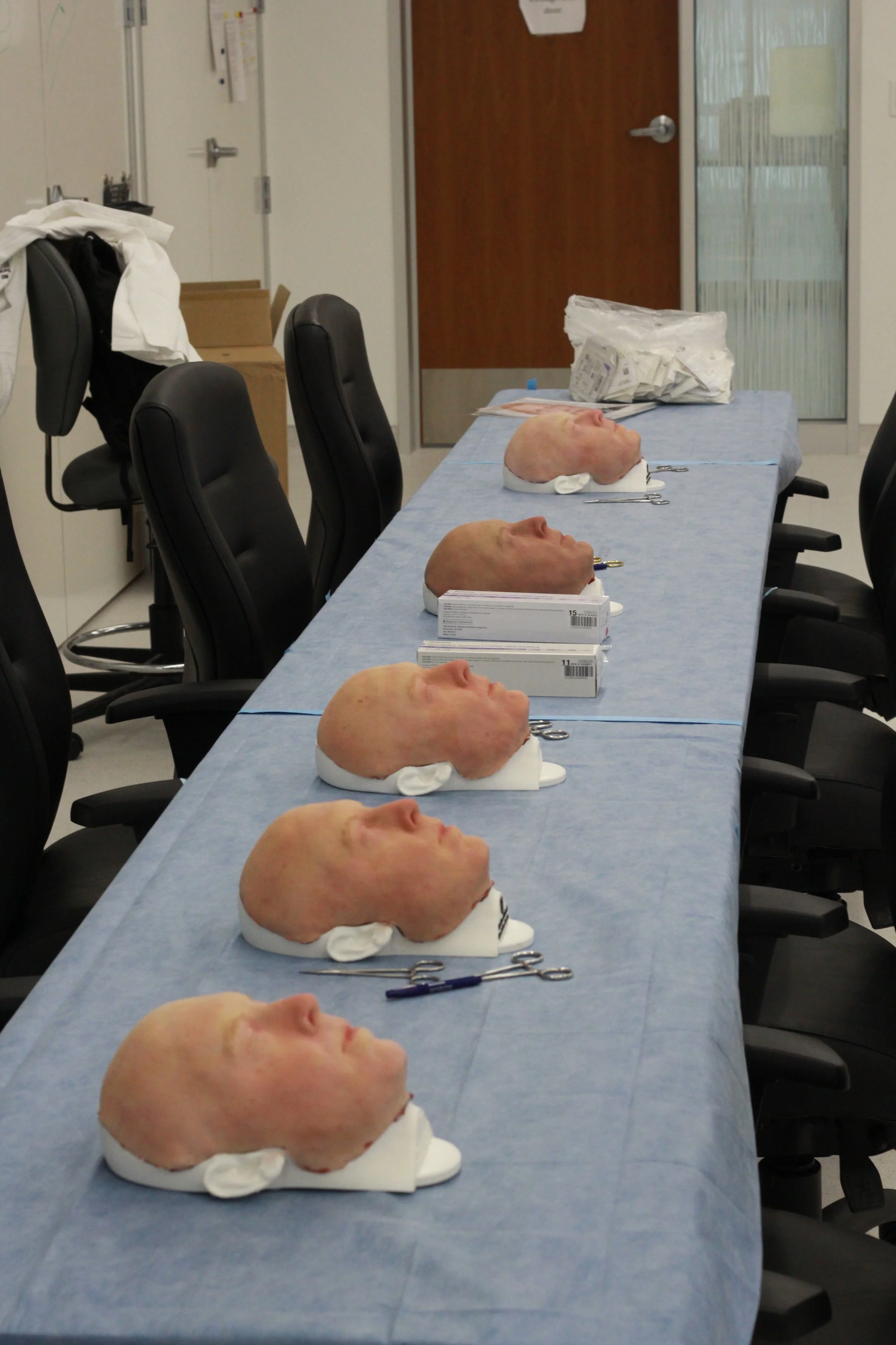
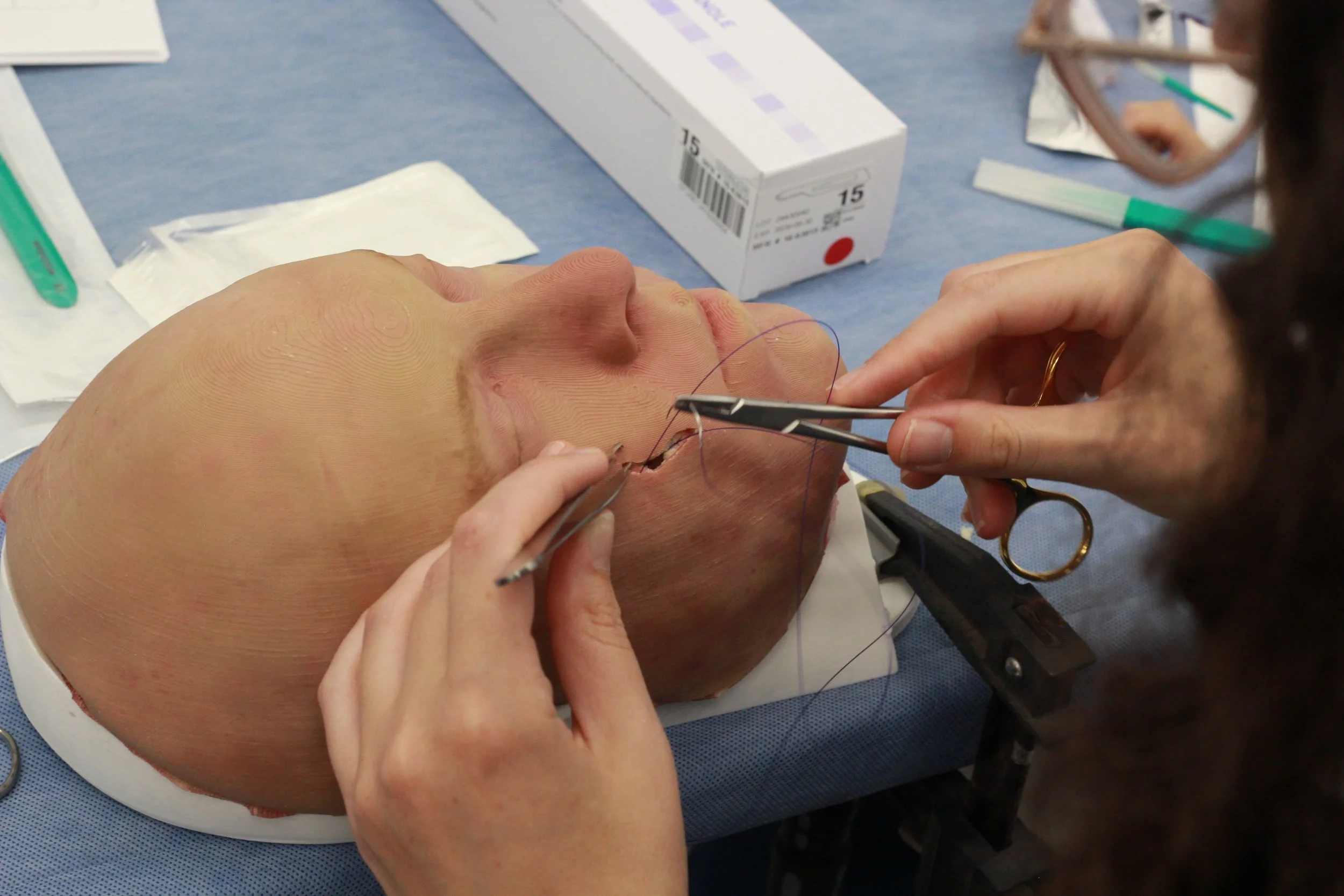
Cutaneous reconstruction using local flaps requires precise planning and experience. Traditionally, training opportunities are provided by using cadavers or other animal skin substiutes (such as pig’s feet). Or learning is through supervised clinical encounters, which is limited by circumstance. Thus, the need for high-fidelity, cost-effective simulation is required to provide safe, on-demand education. We collaborated with the University of Missouri Sheldon Simulation Center and Damon Coyle to design and mass-produce a facial flap task trainer for residents that accurately replicates the anatomical, mechanical, and suturing properties of human facial tissue. Damon is a fantastic artist who has created various simulators for the University of Missouri School of Medicine. For a fascinating glimpse into his work, this link is worth a browse: https://showme.missouri.edu/2022/parts-doctor-parts-artist/
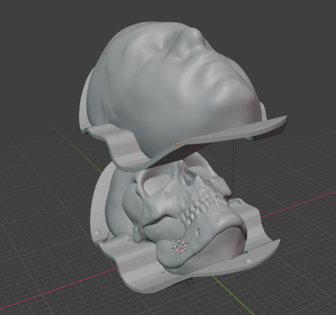
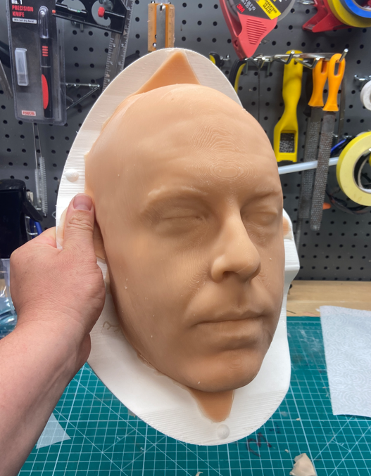
High-resolution 3D scans of a living face gave the team a precise blueprint — every contour and surface captured in sharp detail. From that digital map, designers sculpted and separated the data into parts, then turned those pieces into a multi-part mold system built to recreate the layers beneath the skin.
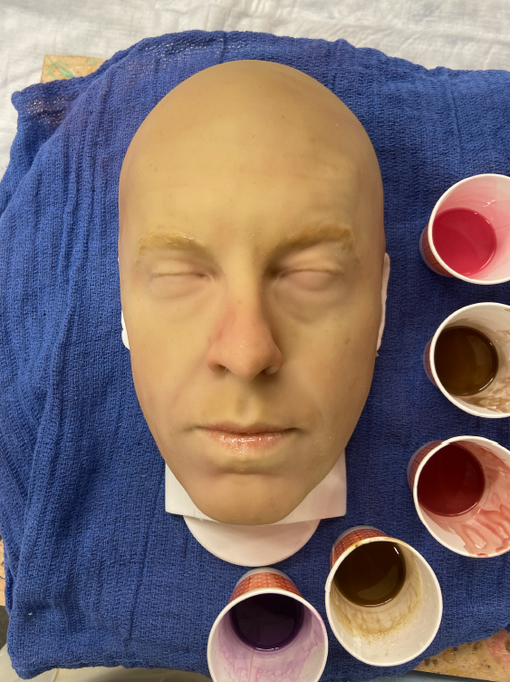
The result is a lifelike facial model made up of four distinct layers: a tough, tear-resistant outer epidermis; a springy dermis; a soft, compliant fat layer; and a fibrous muscle layer. Craftspeople used ultra-soft platinum-cured silicone blends, fine-tuning additives so each layer felt and moved like the real tissue it mimics. The bulk and mobility of the subcutaneous layer came from a mix of silicone foam and fluid-modified silicone, while embedded mesh gave the skin extra tear resistance and made suturing behave realistically. Molds were 3D printed, the layers cast in sequence, then the assembled faces were hand-finished with careful painting and texture work and mounted on a printed skull base to complete the effect.
The finished trainer was put to work in an ENT bootcamp, where residents practiced flap planning, incision techniques, tissue handling and closure. Post-session surveys showed the model scored well for realistic anatomy, effective skill replication and usefulness as a teaching tool. The design was also featured at the “Sim Tank” event during the American Academy of Otolaryngology—Head and Neck Surgery Annual Meeting 2026.
Affordable and reproducible, this facial analogue offers a practical, high-fidelity option for surgical training.